UTC’s electron beam microscope (latest model)
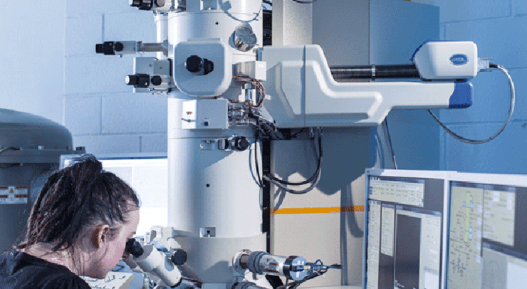
UTC’s acquisition of an electron beam microscope was supported financially by the Picardie Regional authorities under the heading of “structuring equipment” in the State-Region Investment Programme (CPER) 2007–2013. François Oudet, head of co-respondent for equipment purchases under the CPER provisions, tells us more about this 1.3 Meuro instrument.
What are the characteristics of this electron beam microscope?
This model is a TEM/STEM type (Transmission Electron Microscope – Scanning Transmission Electron Microscope) with a field generating cannon. Nominal acceleration is 200 kV. Ultimate resolution is 0.1 nm (in the TEM mode) and 0.2 nm (STEM). It is fitted with a series of devices: two cameras to observe the electron beam (one is a wide angle camera, use mainly to observe living cells and a high resolution camera to observe crystal structures; an electron detector under clear air transmission conditions; an annular electron detector for dark field transmission conditions to observe atomic number contrast levels; and also an X‑ray spectrometer to carry out chemical analyses and produce element mappings of the sample components.
And what does an instrument like this contribute to UTC research?
The microscope is used to characterize matter down to sub-nanometric scale, both from a structural (matter organization) and chemical (composition) point of view. The electron beam microscope has now become a classic tool to characterize and to observe material samples, whether they are metals, ceramic or composites. Thus, all and any ITC research team with investigations of this nature can benefit from the microscope, with the proviso of correctly preparing the samples. Typical applications are characterization of plant tissues, biomimic membranes, divided and nano-materials, agents that interact externally with biological milieus and cell/surface interactions (bio-materials), characterization of catalysts, of materials for surgery (and the health sector in general), nano-metric bone, cell and material structures (biocompatibilities of prosthetic implants, etc.).
What investigations are scheduled today at the electron microscope facility?
Currently we are examining mainly polymer nano-encapsulated particles for use in biological applications as fluorescent markers. A scientific article has already been published on these results: Versatile Synthetic Strategy for Coating Upconverting Nanoparticles with Polymer Shells through Localized Photopolymerization, Using the Particles as Internal Light Sources (Selim Beyazit, Serena Ambrosini, Nataliya Marchyk, Emilia Palo, Vishal Kale, Tero Soukka, Bernadette Tse Sum Bui and Karsten Haupt, Angew. Chem. Int. Ed. 2014, 53, 1 – 6). In this context, the investigation looked at both the morphology of the objects (shape, size, phase distribution) and crystallization. Another theme associates process engineering and looks at the characterization of complex natural oxides in acicular materials. In this case, we observe notably the electron diffractions and use X‑ray spectrometry to complement direct observation of the crystal structures.
How and why do the Picardie Regional authorities support this piece of equipment?
Picardie indeed has strongly supported the projects conforming its high level of commitment in the “structural equipment” category of the CPER 2007–2013, with 1.25 Meuros out of 1.3 Meuros (before tax), plus 50 000 euros from the FEDER (European Regional development Fund). The project presentation qualified the microscope as a priority, transversal acquisition to be used in UTC themes that contribute to the Picardie Region’s competitivity (agro-resources, health and material sciences and engineering) and which tie in closely with research conducted by 4 of UTC’s research units (GEC, Roberval BMBI and TIMR). The UTC Research Directorate present the acquisition project to the CPER ministerial authorities and to the FEDER offices in Brussels, and above all to the Picardie Region, insisting on the scientific importance of possessing such a facility and the need for having such a microscope in terms of regional thematics, and sharing of its use among the various UTC research units. The latter point was highly appreciated by the CEPER specialists. Continuous exchanges with Virginie Delaporte (in the Picardie Region Research and Innovation Services) enables us to implement our procurement policy for high level modern analytical tools destined to be shared among laboratories and others. Given the price tag the microscope has become the ‘emblematic’ tool but there are also other highly important equipment units such as the atomic force microscope, a confocal laser microscope, an NMR spectrometer and soon, a rehabilitated ‘environmental’ electron microscope.