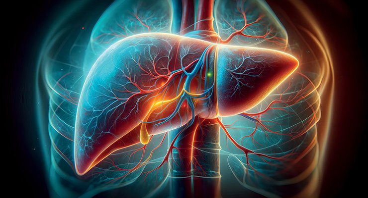
The hepatogram: measuring liver rigidity
Sabine Bensamoun is a research director at the CNRS and works in the CNRS/BMBI joint research unit at UTC. She is also a research associate at the Mayo Clinic (Rochester, USA) and a member of the CNRS National Committee (CoNRS). With Dr Fabrice Charleux of ACRIM, she took part in the international liver study that led to the hepatogram.
In practical terms? «Until now, to analyse the level of rigidity of the liver, a doctor would prescribe an examination of the organ using the magnetic resonance elastography (MRE) technique. From 2024, as with an electrocardiogram, doctors will be able to prescribe a hepatogram, a name that will be approved in 2023, for a specific examination of the liver. It’s a quick, non-invasive test that provides a more accurate diagnosis. MRI gives a purely anatomical image of the organ analysed, whereas MRE gives us an image that indicates the degree of rigidity of the tissue analysed,» explains Sabine Bensamoun.
The story of MRE began when she returned to France after her post-doc at the Mayo Clinic, a world reference in medical research. She joined the CNRS and continued her collaboration with the Mayo Clinic, where a module had been developed which, coupled with MRI (magnetic resonance imaging), aims to better characterise the mechanical or functional properties of soft organs, including the liver. This is known as magnetic resonance elastography (MRE). This was not available in France when it was introduced in 2006. According to Sabine Bensamoun, what does this technique offer compared with other imaging techniques? In particular, it enables us to obtain a better diagnosis of the severity of pathologies, improve patient follow-up and, lastly, personalised treatments, etc.». All that remained was to validate the MRE clinically..
The UTC is one of ten research centres selected by the Mayo Clinic worldwide, and the only one in France, to benefit from this module. It was still to be coupled it to an MRI machine. «I got in touch with Dr Fabrice Charleux, a radiologist at ACRIM and told him about my plans; he was interested, particularly by the research aspect. At the time, he was working on General Electric machines, which proved opportune and useful because the module developed by the Mayo Clinic, which was only a prototype, only worked with this type of machine. For more than 10 years, I continued to improve the protocol I had first developed at the Mayo Clinic on muscle tissues, in particular for the disorder known as Duchenne muscular dystrophy and muscle ageing», she points out.
After years of research, the data collected from all over the world was analysed at the Mayo Clinic. For all the parties involved, including UTC and ACRIM, the gamble paid off. The result is the MRE, a non-invasive diagnostic tool for analysing the entire liver to establish the stage of fibrosis.
And what is your interest in the human liver? «In fact, the Mayo Clinic continued to improve the module for future commercialisation. At the time, they were working extensively on the liver organ and wanted to have broader sources of data. To do this, several research centres had to be equipped with this new module. As far as we were concerned, we started working on the liver, but with the former module,» she says.
How does an MRE work? «If we take the liver as an example, we can see that the more diseased it is, the more rigid it becomes. Thanks to MRE, we can quantify the organ’s rigidity, with stages ranging from one to four, the highest indicating liver cirrhosis (4). The module takes the form of a box fitted with a speaker that sends low-frequency air pressure through a stimulator that is placed on the tissue to be analysed. The movement of this low-frequency vibration is then monitored and, depending on the speed at which the wave propagates through the organ concerned, the degree of severity of the pathology can be estimated. It’s a real alternative to biopsy,» describes Sabine Bensamoun.
Of all the soft tissues, the liver is currently the one where MRE is performed as a routine clinical procedure. In the United States, more than 100 000 examinations, now known as hepatograms, were carried out in 2023, and more than 2,300 newgeneration MREs have been installed worldwide, including the one at ACRIM.
What’s next? «Our expertise in fibrous pathological tissues (muscle, liver) was invoked during the COVID-19 pandemic. We are already working on a clinical protocol applied to lung tissue,» she concludes.