Improving the diagnosis and treatment of biliary diseases

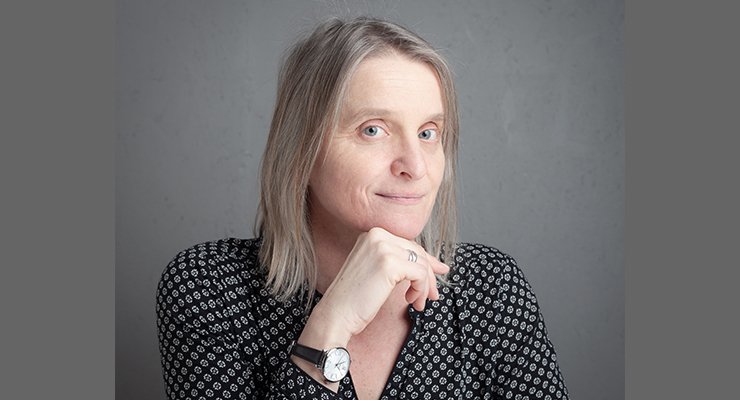
Isabelle Claude is a lecturer specialist in medical image processing, particularly in multi-modality image segmentation, currently working on the MAAGIE project funded by the ANR, the aim of which is to improve the diagnosis and treatment of biliary diseases.
Isabelle Claude works with magnetic resonance imaging as well as computed tomography and ultrasonics. This leads her, depending on the clinical request submitted to her, to develop specific tools.
Launched in January 2025 for a period of four years, MAAGIE brings together three laboratories: UTC-BMBI, ISIR and LIP6 of Sorbonne University and two AP-HP hospitals, Saint-Antoine Hospital in Paris and Henri-Mondor Hospital in Créteil. The aim of the project is to improve the diagnosis and treatment of lithiasic and tumorous diseases of the bile ducts, both of which prevent bile from flowing, with potentially dramatic consequences for the patient.
The idea behind this project? “It’s about giving gastroenterologists the benefit of the latest technological developments in both robotics and digital technology, as has been the case in the vascular and cardiac fields. The aim is to improve the success rate of endoscopic retrograde cholangiopancreatography (ERCP). A complicated intervention since it involves inserting an endoscope through the patient’s mouth, passing it through the stomach, then finding the papilla, an opening in the duodenum through which instruments are inserted. These will be passed up into the bile ducts either to remove stones or to insert stents,” explains Isabelle Claude. This double endoluminal access causes 5 to 10% of the procedure to fail.
What are the solutions to reduce this failure rate? As part of his thesis, Abdelhadi Essamlali adapted a U‑Net model, a tool based on convolutional neural networks, to the problem of image segmentation and then 3D reconstruction of the biliary tree. A tool validated by clinical partners. “The first step is to reconstruct the bile ducts in 3D using 2D images (MRI, scanner) of the patient before the operation. This allows the practitioner to better follow the patient’s anatomy and thus better plan the ERCP. Then, in the preoperative phase, the 3D reconstruction is merged with the planar fluoroscopy images to help the surgeon find the right duct to treat,” concludes Isabelle Claude.
MSD


